Generating Regenerative Promise

Stem cell research has tremendous potential to create regenerative cells and treat disease
Story by Richard Asa
SCIENTISTS STUDYING STEM CELLS at the College of Medicine are generating a great deal of interest. Why? Because of the burgeoning potential to use these undifferentiated cells as the basis of regenerative treatments for a plethora of conditions.
They work with totipotent and pluripotent stem cells, which are able to differentiate into many functional cell types including various neurons, heart cells and lung cells. This allows for the regeneration and replacement of cells in patients.
For example, last year College of Medicine researchers identified a molecular mechanism that directs pluripotent stem cells to mature into endothelial cells—the specialized cells that line blood vessels and maintain healthy blood flow.
Understanding how that process unfolds could help scientists more efficiently convert stem cells into endothelial cells for use in tissue repair, or for engineering blood vessels to build new organs and tissues.
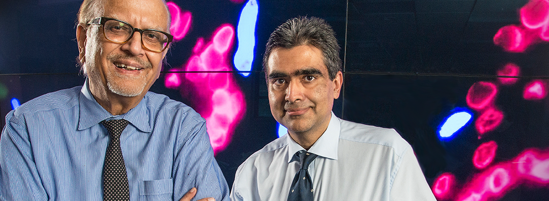
The research team led by Asrar Malik, PhD, Schweppe Family distinguished professor and head of pharmacology at the College of Medicine’s Chicago campus, studied the enzymes that regulate how DNA is transcribed into RNA. The research team identified two such epigenetic enzymes controlling how stem cells turn into blood vessel lining cells. Understanding these epigenetic switches is an important step in unraveling the gene networks that determine the fate of stem cells.
Malik recognizes the fundamental importance of regenerative therapies in the decades to come. That led to his decision to develop an extensive regenerative biology and medicine program at the College of Medicine’s Chicago campus.
His own research has focused on repairing blood vessels in lungs after severe infection, but he also actively works with teams of researchers who are studying regeneration and stem cells in the heart and other tissues.
“Our goal is to understand how to repair and regenerate diseased blood vessels as well as grow new blood vessels using stem cells,” says Jalees Rehman, MD, associate professor of medicine (cardiology) and pharmacology, who previously taught at the University of Chicago. “Blood vessels are necessary to maintain the health of all organs in the body because they supply the necessary nutrients and oxygen.
“High cholesterol levels, diabetes, high blood pressure and inflammation are examples of factors that can damage or even destroy endothelial cells—the important cells which line our blood vessels,” he adds. “Patients with damaged blood vessels unfortunately suffer terrible consequences of the diminished blood flow, such as heart attacks and strokes.”
Once blood vessels are diseased, treatments that could reverse the damage by supplying newer, healthy blood-vessel endothelial cells will be needed. Regenerative biology provides the tools to use stem cells to restore healthy blood vessel function, Rehman says.
Another exciting possibility offered by stem cells is their potential ability to serve as the building blocks to engineer entire organs. The demand for organ transplants is far outstripping the supply of donor organs, so biomedical researchers including Rehman and Malik have turned their attention to using stem cells to build blood vessel networks that can be used for new organs and tissues that could be implanted in patients.
These investigators believe that a better understanding of the molecular and cellular mechanisms are needed before therapeutic repair of blood vessels and the engineering of new tissues and organs for transplants can be implemented.
Other important stem-cell-related research at the COM includes:
- Using zebrafish to study the dynamic interactions between blood stem cells and their niche micro-environment. The blood system in zebrafish is remarkably similar to that of humans and allows the advantage of directly watching blood stem cells and how they behave in real time. By treating zebrafish with drugs, researchers can rapidly screen compounds that boost blood stem cells. These drugs have the potential to translate to clinical trials for patients in need of stem cell transplantation.“I believe this will allow us to rapidly improve stem cell transplants in the clinic, and give patients a better chance of survival,” says assistant professor of pharmacology Owen Tamplin, PhD.
- Providing a renewable source of blood for patients in need of recurrent transfusions and transplantations, which is the case with immune-compromised patients or those suffering from various forms of leukemia. The major research focus is the expansion and self-renewal of hematopoietic stem cells.We study blood regeneration by investigating signaling pathways that regulate stem cell decisions responsible for generation of blood cells,” says assistant professor Kosta Pajcini, PhD. “One means by which blood cells decide their fates is by instructions they receive from their neighboring cells.” For example, in the bone marrow, most of the blood stem cells are told to remain as stem cells by signals that they receive from their niche cells (unless there is a need for replenishment of blood, at which point the stem cells differentiate into mature blood cells).Pajcini adds that he wants to use his knowledge of cell signaling to create an environment in a laboratory incubator that would allow for indefinite expansion of blood stem cells. He could then test the ability of cultured stem cells to reconstitute the hematopoietic system in a transplantation setting.
- Examining how stem cells interact with their physical environments such as fluid shear and the mechanics of the extracellular matrix. An understanding of this dynamic would help researchers control how stem cells are distributed in the body after injection and how they exert their function at target sites. By incorporating physical cues, the efficacy of stem cells can be improved through their ability to secrete soluble factors to program neighboring blood and immune cells.“To achieve these goals, our laboratory offers an interdisciplinary research environment at the interface between medical science and engineering,” says assistant professor of pharmacology Jae-Won Shin, PhD. For instance, his lab develops various approaches in biophysics and engineering, such as micropipetting, atomic force microscopy, microfluidics, and biomaterial design to both manipulate and measure physical forces exerted on stem cells.Such approaches are combined with molecular biology and in vivo animal models not only to understand fundamental mechanisms behind how physical forces regulate different aspects of stem cell functions and develop biophysical approaches to stem cell therapies, he adds.
“The field of stem cell biology has evolved tremendously in the past seven years in our department,” Malik says. “We’ve recruited talented investigators from the best programs nationally, who are driving discovery and adding to our strengths. There are currently seven faculty members whose primary effort and funding relates to stem cell biology. This has been and will be continue to be a concerted effort in the coming years and a major area of focus.
“The intent is to develop nationally ranked program in stem cell biology and regenerative medicine and also provide advanced research training to the future generation,” he adds. “The work is currently supported by a dozen or so highly competitive NIH grants and also there is a growing effort to seek out private support.”
Stem Cell in Peoria and Rockford
THE RESEARCH IN CHICAGO IS COMPLEMENTED by stem cell work at the College of Medicine Peoria and Rockford campuses. In Rockford, the head of the department of biomedical sciences, Ramaswamy Kalyanasundaram, PhD, recently started a regenerative and disability laboratory to make advances in finding cures or preventive measures for disability-related conditions. The lab was made possible with a gift from the Blazer Foundation with matching funds from the COM.
The lab is headed by two researchers who focus on differentiating stem cells into motor neurons and using stem cells to improve the integration of metal implants into the surrounding bone tissue. Xue-Jun Li, PhD, will work with embryonic stem cells and patient-specific induced pluripotent cells to create motor neurons that can eventually be transplanted into patients with spinal muscular atrophy and hereditary spastic paraplegias. The stem cells being used are derived from patients with those diseases.
Li’s group also will build high-throughput and high content drug screening systems to identify therapeutic agents for slowing down motor neuron degeneration and promoting nerve regeneration. She will be collaborating with scientists at the National Institutes of Health, the University of Wisconsin-Madison and the University of Connecticut.
“Degeneration of motor neurons, large projection neurons controlling the movement of muscles, underlies many debilitating diseases,” she says. “One focus is to direct human pluripotent stem cells into both upper and lower motor neurons. Our lab has established a unique paradigm to efficiently generate spinal motor neurons from human pluripotent stem cells.
The other type of motor neuron, upper, is specified by a different mechanism. Li wants to establish a system to generate currently unavailable human upper motor neurons and build 3-D neural tissue co-culture models to study the connections between upper and lower motor neurons. Her research also focuses on using human pluripotent stem cells to model motor neuron diseases and spinal cord injury. The lab has already successfully established human stem cell models for spinal muscular atrophy and hereditary spastic paraplegias.
Mathew Thoppil-Mathew, PhD, will be developing new materials and suitable surface design modifications for hip, knee and jaw replacement and dental implants. His research will investigate the various causes of joint corrosion after replacement and develop technologies to minimize the wear and tear on the joint.
Mathew is an active research faculty at the department of restorative dentistry, UIC College of Dentistry, and the department of orthopaedic surgery at Rush University Medical Center, Chicago. He is also collaborating with scientists in Brazil, Germany, United Kingdom, Portugal and South Africa. Both Li and Thoppil-Mathew have joint appointments in the department of bioengineering under Thomas Royston, PhD, department head.
“The success of any metal implants is heavily dependent on their integration with the surrounding bone tissue,” Thoppil-Mathew says. “Damage to the bone tissue can result in the failure of metal implants. So, our research will focus on ways to improve bone tissue regeneration around the implants, and our lab will study the metal surface, cell, protein interactions during corrosionwear and develop approaches to minimize the damage.”
In Peoria, a research team is investigating ways to use stem cells to prevent further damage and facilitate recovery from stroke, a leading cause of death and a leading cause of serious long-term disability. About 795,000 people have a stroke each year in the U.S., the CDC says, and about 130,000 Americans die—that’s one in every 20 deaths.
Krisha Kumar Veeravalli recently received the Sudhir Gupta Young Scientist Award from the National Association of Scientists of Indian Origin in America, recognizing his contributions in the field.
Krishna Kumar Veeravalli, PhD, and his team, who have worked on stroke research since 2011, are trying to advance pharmacotherapy that will prevent further brain damage and improve functional recovery through regeneration after a stroke. To that end, he has continually expanded a study showing that mesenchymal stem cells derived from human umbilical cord blood can prevent the ongoing brain cell death after stroke.
“We’ve seen the potential that stem cells have for providing neuroprotection and recovery after stroke and spinal cord injury,” he says. “The main objective of our current studies is to determine how age and sex differences affect treatment outcomes after stem cell therapy. This will help validate stem cell use as a benefit for both sexes including post-menopausal females.”
Veeravalli says his team’s research has suggested that the combination of stem cell treatment and gene silencing approach, which could target multiple molecules/pathways, could be the key to obtain a substantial therapeutic benefit after stroke.
“It is very important to target several key molecules simultaneously, which can work by multiple mechanisms to control the early and delayed brain injury after stroke” he says. “Our novel approach would adopt preclinical testing, advance the understanding of the pathophysiology of stroke and make it possible to translate it from bench to bedside.”
Ultimately, Malik sees exciting trends in biomedical research with great potential for translational research in the area of stem cell biology, which will become a pillar of 21st century medicine. “We’ve established a significant program with much room for growth, according to our external consultants. It is a rapidly changing field, and it will take time and a lot of hard work, smart energetic people and resources, but also a great deal of good luck and serendipity. We’re answering fundamental questions that are central to many aspects of biology. But it will be very much worth the effort.”